
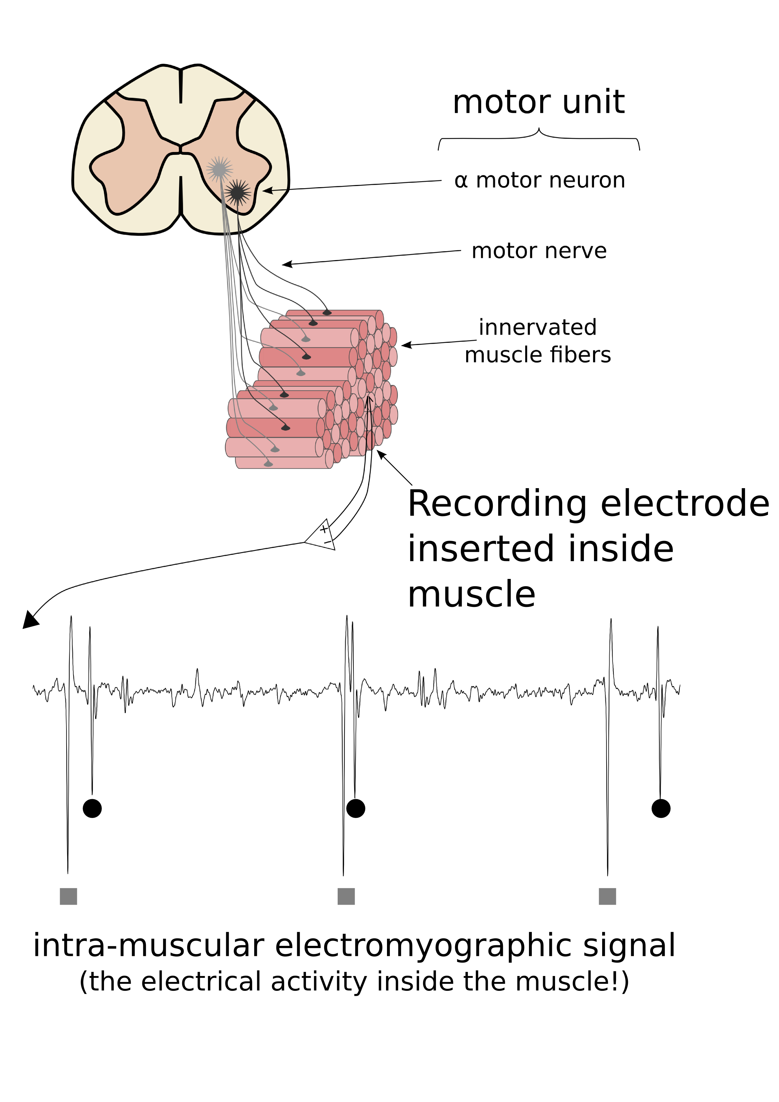
Motor units consist of a motoneuron located in the spinal cord, a peripheral axon that goes from the spinal cord to the muscle, and the various muscle fibres that are innervated by the motoneuron. When a recording electrode is positioned inside a muscle, the simultaneous activation of the muscle fibres innervated by a single motoneuron result in an electrical potential that has a characteristic shape. These shapes (motor unit action potentials) can be analysed and the activity from a single motoneuron located in the spinal cord can be determined.