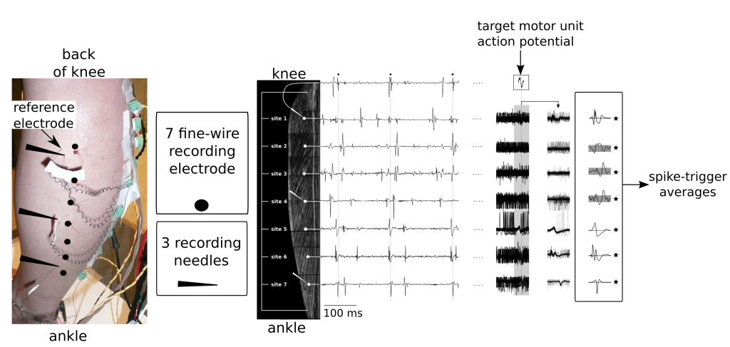
On the left we see the (scary) experimental set-up with the 3 micro-electrodes and 7 fine-wire electrodes inserted along the length of the medial gastrocnemius muscle. By recording the activity at various locations along the length of the muscle, and then using the discharge times of a target motor unit (Middle panel: small black circles on the top trace)to trigger the activity of the other recordings, spike-triggered averages could be computed. In this example, the target motor unit likely had muscle fibres along the full length of the medial gastrocnemius.