Body mass and body size increase during growth. Muscles must adapt during growth to deal with these changes. However, little is known about the modifications in the three dimensional geometry (e.g. cross-sectional area, fibre length) of developing muscles (Bénard et al. 2011; Böl et al. 2017). Knowledge about the structural changes in muscle morphology is of great interest in medicine, biomechanics, and modeling. For example, in neuromuscular disorders such as cerebral palsy, muscle growth is hampered and joint mobility is thus limited. Young patients often undergo surgical muscle lengthening to improve their gait mechanics, and improving treatment of these muscles requires detailed insight into how their geometry develops during growth. Also, data on muscle morphology can be used to develop and validate muscle growth models.
WHAT DID WE FIND?
The present study (Siebert et al. 2017) analyzed growth-related changes in muscle fascicle lengths, pennation angles, tendon lengths, muscle cross-sectional areas, and aponeurosis area of the gastrocnemius (lateralis and medialis), flexor digitorum longus, and tibialis anterior in the rabbit.
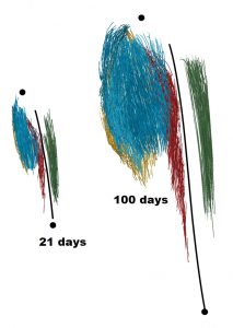
We found an almost linear increase over time in most of the geometrical parameters. Gastrocnemius lateralis and medialis showed very similar growth characteristics. In general, the aponeuroses of all muscles exhibited lower growth rates in width than in length. Furthermore, aponeurosis areas were larger than physiological cross-sectional areas. There were almost no changes in fascicle lengths with increasing age for gastrocnemius (lateralis and medialis) and flexor digitorum longus. In contrast, there was a clear increase in fascicle length of tibialis anterior. Pennation angles of tibialis anterior and flexor digitorum longus remained almost unchanged but increased for gastrocnemius lateralis from about 13° to 24° during growth.

Lateral view of 3D rabbit shank muscle at 21 and 100 days of age. Blue is gastrocnemius lateralis. Yellow is gastrocnemius medialis. Green is tibialis anterior. Red is flexor digitorum longus.
For all muscles, the tendon-muscle fascicle length ratio (tendon length divided by fascicle length) changed during growth. We found an increase in this ratio by a factor of 2 for gastrocnemius (lateralis and medialis) and flexor digitorum longus, but a slight decrease for tibialis anterior. The increase in the ratio might indicate increasing importance of economical locomotion during growth as longer tendons (in relation to the muscle fibers) enable a higher strain energy storage capacity with increasing body mass.
SIGNIFICANCE AND IMPLICATIONS
The study’s results provide new information about changes in three-dimensional muscle architecture and aponeurosis shape during growth. The results help to improve our understanding of muscle growth processes, and they might be important in diagnoses of neuromuscular disorders. Moreover, the results might be relevant for understanding changes in muscle energetics and economy of locomotion during growth.
PUBLICATION REFERENCE
Siebert T, Tomalka A, Stutzig N, Leichsenring K, Böl M. Changes in three-dimensional muscle structure of rabbit gastrocnemius, flexor digitorum longus, and tibialis anterior during growth. J Mech Behav Biomed Mater, 74: 507-519, 2017.
If you cannot access the paper, please click here to request a copy.
KEY REFERENCES
Bénard MR, Harlaar J, Becher JG, Huijing PA, Jaspers RT. Effects of growth on geometry of gastrocnemius muscle in children: a three-dimensional ultrasound analysis. J Anat 219: 388–402, 2011.
Böl M, Leichsenring K, Siebert T. Effects of growth on muscle, tendon and aponeurosis tissues in rabbit shank musculature. Anat Rec 300: 1123-1136, 2017.
AUTHOR BIO
 Tobias Siebert is a Professor of motion and exercise science at the University of Stuttgart in Germany. His main focus in research is modeling and experimental investigations of the neuromuscular system. You can learn more about Tobias and his research here.
